


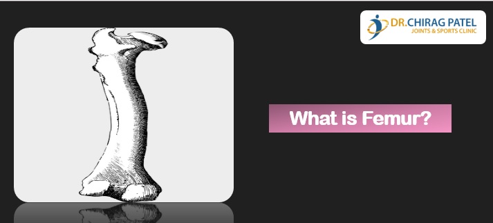
The femur is the only bone in the thigh. It is the longest bone in the body. The main function of the femur is to transmit forces from the leg to the hip joint. It acts as the site of origin and attachment of many muscles and ligaments, and can be divided into three areas; proximal, shaft and distal.
The proximal area of the femur forms the hip joint with the pelvis. It consists of a head and neck, and two bony processes called trochanters. There are also two bony ridges connecting the two trochanters.
- Head– Articulates with the acetabulum of the pelvis to form the hip joint. It has a smooth surface with a depression on the medial aspect; for the attachment of the ligament of head of femur
- Neck– Connects the head of the femur with the shaft. It is cylindrical, projecting in a superior and medial direction – this angle of projection allows for an increased range of movement at the hip joint.
- Greater trochanter– A projection of bone that originates from the anterior aspect, just lateral to the neck. It is angled superiorly and posteriorly, and can be found on both the anterior and posterior sides of the femur is the site of attachment for many of the muscles in the gluteal region, such as gluteus medius, gluteus minimus and piriformis.
- Lesser trochanteris smaller than the greater trochanter. It projects from the posteromedial side of the femur, just inferior to the neck-shaft junction is the site of attachment for the psoas major and iliacus muscles.
- Intertrochanteric crest is a ridge of bone that connects the two trochanters together. It is located on the posterior surface of the femur.
The shaft descends in a slight medial direction. This brings the knees closer to the body’s center of gravity, increasing stability. On the posterior surface of the femoral shaft, there are roughened ridges of bone, these are called the linea aspera (Latin for rough line). Proximally, the medial border of the Linea Aspera becomes the pectineal line. The lateral border becomes the gluteal tuberosity, where the gluteus maximus attaches. Distally, the linea aspera widens and forms the floor of the popliteal fossa, the medial and lateral borders form the medial and lateral supracondylar lines. The medial supracondylar line stops at the adductor tubercle, where the adductor magnus attaches.
The distal end of the femur is characterized by the presence of the medial and lateral condyles, which articulate with the tibia and patella, forming the knee joint. Medial and lateral condyles are rounded areas at the end of the femur. The posterior and inferior surfaces articulate with the tibia (leg/shin bone) and menisci of the knee, while the anterior surface articulates with the patella or the knee cap. Medial and lateral epicondyles are bony elevations on the non-articular areas of the condyles. They are the area of attachment of some muscles and the collateral ligaments of the knee joint. Intercondylar fossa is a depression found on the posterior surface of the femur, it lies in between the two condyles. It contains two facets for attachment of internal (anterior and posterior cruciate) knee ligaments.
Get in touch with Dr Chirag Patel at Sunshine Global Hospital Surat, for all Knee Injury Treatment. Contact +917574887777.